Fluorescence microscopy is a technique whereby fluorescent substances are examined in a microscope, It has a number of advantages over other forms of microscopy, offering high sensitivity and specificity, In fluorescence microscopy, the specimen is illuminated excited with light of a relatively short wavelength, usually blue or ultraviolet UV,
fluorescence-microscopy-imaging GitHub Topics GitHub
Using live cell confocal microscopy and flow cytometry we show that the tC O-labeled mRNA is efficiently translated into H2B:GFP inside human cells Hence we not only develop the use of fluorescent base analogue labeling of nucleic acids in live-cell microscopy but also importantly show that the resulting transcript is translated into the correct protein Moreover, the spectral properties of …
Cited by : 2
Fluorescence Microscopy and Advanced Imaging
Fluorescence Imaging
Tissues, cells, and the smaller structures inside cells organelles are mostly water and are therefore transparent, Imaging tiny see-through bags of water results in images that don’t contain a lot of information, and in microscopy, it is vital to have some sort of contrast or stain that will give areas of the sample color and make them far easier to see, In addition, what if you only want to image some of the smaller str…
Fluorescence Microscopy – an overview
Fluorescence microscopy is a popular imaging technique both for its relatively noninvasive properties and its ability to simultaneously image multiple species in living cellular specimens, Images are formed by the simultaneous observation of many fluorescing molecules to visualize cellular structures and processes, In conventional far-field fluorescence microscopy, the spatial resolution is diffraction …
Fluorescence lifetime imaging microscopy FLIM is a method for measuring fluorophore lifetimes with microscopic spatial resolution providing a useful tool for cell biologists to detect visualize and investigate structure and function of biological systems, In this chapter, we begin by introducin …
Cited by : 31
· Fluorescence microscopy tensor imaging representations of the cardiac microvascularity We then used our method to interrogate the cardiac microvasculature and obtain structural tractographic
Cited by : 20
Stealth Fluorescence Labeling for Live Microscopy Imaging
to acquire images in fluorescence microscopy Combined manipulations The Inscoper user interface is natively designed so that all microscopy manipulations SPIM, FRAP, high-content screening, etc, …
Fluorescence Microscopy & Cell Imaging Microscopy is at the heart of understanding tissue architecture, cell structure and dynamics, as well as molecular function, Fluorescence microscopy is routinely used to determine spatial and topological information about cells and tissues,
Fluorescence Microscopy, KeyLight delivers intense, broad-spectrum UV and visible wavelengths for a wide variety of colors between 340nm and 760nm, These compact light sources support 3, 4, 5, 6 or 7 channel systems with customizable wavelengths and UV intensity for greater flexibility depending on the application requirements, The product form factor and control options are designed specifically to …
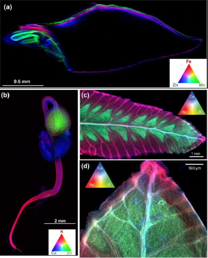
Fluorescence microscopy: biological applications and
· Fluorescence microscopy: biological applications and imaging methods – An Introduction Fluorescence microscopy is an enormously powerful tool for investigations in the biological field Fluorescence microscopy is more than “just making colorful images in one two three or even more colors”, Fluorescence techniques place numerous benefits in the hands of researchers wishing to …
Fluorescence microscopy tensor imaging representations for
fluorescence microscopy imaging
Fluorescence Microscopy & Cell Imaging
Fluorescence Microscopy – an overview
Fluorescence-lifetime imaging microscopy
Overview
Fluorescence imaging
Overview
· GitHub is where people build software More than 65 million people use GitHub to discover fork and contribute to over 200 million projects
Fluorescence lifetime imaging microscopy for quantitative
INSCOPER